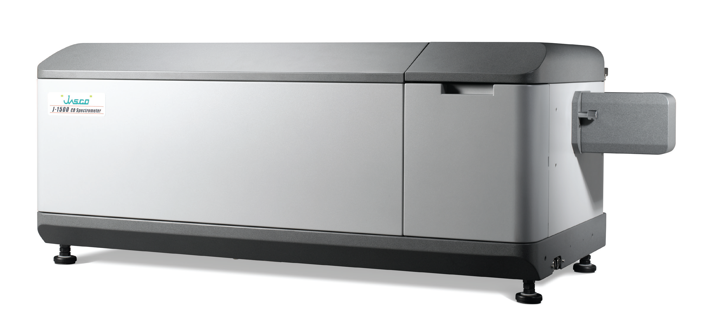
Secondary structure analysis of poly-L-glutamic sodium using titration with dilute sulfuric acid

In the fields of fundamental protein research and pharmaceutical technology, studies into protein structure and peptides models are becoming of increasing interest; the function of proteins is intrinsically linked to their structure, and the structural analysis of proteins and peptide models is important in determining their bioactivity.
NMR and X-ray crystal structure analysis are both very effective methods in the elucidation of protein structure, but the requirement for large amounts of sample and equipment costs can be prohibitive for routine analysis.
By comparison, analysis using circular dichroism is very straightforward and can be done with small amounts of sample. These features make CD a useful tool for the estimation of secondary structure of proteins and peptides, analyzing the conformational changes caused by pH, temperature, and ligand binding.
In structural analysis using CD measurement, the abundance ratio of secondary structure motifs in proteins and peptides can be estimated using a least-square method with reference spectra including α-helix, β-sheet, turn, and random structure. The JWSSE-513 protein secondary structure analysis program uses a Classical Least Squares (CLS) method, which includes the reference spectra of Yang and Reed. Yang’s reference spectra are extracted from CD spectra of proteins, and are best suited to protein secondary structure analysis. Reed’s reference spectra are extracted from the CD spectra of peptides, and are suited to the secondary structure analysis of peptides; because of lesser effects on CD caused by the side chains of aromatic amino acids often found in proteins. For the secondary structure estimation of peptides, the JWSSE-513 protein secondary structure analysis program using Reed’s reference spectra is extremely effective.
The data shown in this application note is an example of CD spectral change in poly-L-glutamic acid sodium solution with titration with dilute sulfuric acid. The changes in secondary structure are reported.
